
Mouse Secondary Antibodies

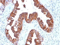
Biotium Cytokeratin 7 (Glandular and Transitional Epithelial Marker) (rOV-TL12/30), CF740 conjugate, 0.1mg/mL
This antibody recognizes an intermediate filament protein (IFP) of 55 kDa, which is identified as cytokeratin 7. This MAb is highly specific to cytokeratin 7 and shows no cross-reaction with other IFPs. Cytokeratin 7 is a basic cytokeratin, which is found in most glandular and transitional epithelia but not in the stratified squamous epithelia. Keratin 7 is expressed in the epithelial cells of ovary, lung, and breast but not of colon, prostate, or gastrointestinal tract. This MAb is highly useful in distinguishing ovarian carcinomas (keratin 7 ) from colon carcinomas (keratin 7-).Primary antibodies are available purified, or with a selection of fluorescent CF Dyes and other labels. CF Dyes offer exceptional brightness and photostability. Note: Conjugates of blue fluorescent dyes like CF405S and CF405M are not recommended for detecting low abundance targets, because blue dyes have lower fluorescence and can give higher non-specific background than other dye colors.

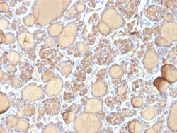
Biotium Thyroglobulin (Thyroidal Cell Marker) (r6E1), Biotin conjugate, 0.1mg/mL
Thyroglobulin is a 660 kDa dimeric pre-protein with mutiple glycosylation sites. It is produced by and processed within the thyroid gland to produce the hormone thyroxine and triiodothyronine. Prior to forming dimers, thyroglobulin monomers undergo conformational maturation in the endoplasmic reticulation. The vast majority of follicular carcinomas of the thyroid will give positive immunoreactivity for anti-thyroglobulin even though sometimes only focally. Poorly differentiated carcinomas of the thyroid are frequently anti-thyroglobulin negative. Adenocarcinomas of other-than-thyroid origin do not react with this antibody. This antibody is useful in identification of thyroid carcinoma of the papillary and follicular types. Presence of thyroglobulin in metastatic lesions establishes the thyroid origin of tumor. Anti-thyroglobulin, combined with anti-calcitonin, can identify medullary carcinomas of the thyroid. Furthermore, anti-thyroglobulin, combined with anti-TTF1, can be a
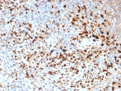
Biotium HSV1 (Herpes Simplex Virus Type I)(N/A), CF740 conjugate, 0.1mg/mL
The antibody reacts with HSV type 1 specific antigen. It is suitable for detection of HSV in human cellular material obtained from superficial lesions or biopsies and for the early identification of HSV in infected tissue cultures. The herpes simplex virus (HSV) (also known as cold sore, night fever or fever blister) is a virus that causes a contagious disease. There are two main types of Herpes Simplex Virus (HSV), 1 and 2. The HSV-1 strain generally appears in the orafacial organs. HSV2 usually resides in the sacral ganglion at the base of the spine. All herpes viruses are morphologically identical: they have a large double-stranded DNA genome and the virion consists of an icosahedral nucleo-capsid, which is surrounded by a lipid bilayer envelope. Primary antibodies are available purified, or with a selection of fluorescent CF Dyes and other labels. CF Dyes offer exceptional brightness and photostability. Note: Conjugates of blue fluorescent dyes like CF405S and CF405M

Biotium Aurora B (Proliferation Marker)(AURKB/1845), CF568 conjugate, 0.1mg/mL
This antibody recognizes a protein of 39 kDa, which is identified as Aurora kinase B (or Aurora B). Aurora B is a serine/threonine protein kinase and component of the chromosomal passenger complex (CPC), a complex that acts as a key regulator of mitosis. It is important for accurate chromosome segregation, cytokinesis, protein localization to the centromere and kinetochore, correct microtubule-kinetochore attachment, and regulation of the mitotic checkpoint. Aurora B forms a tight complex with inner centrosome protein and survivin. Inactivation of any of these proteins causes similar defects in chromosome segregation. A significant overexpression of Aurora B has been found in a variety of human tumors including non-small cell lung carcinoma, astrocytoma, seminoma and carcinomas of the colon, prostate, endometrium and thyroid. The expression level of Aurora B is associated with cell proliferation and prognosis in these tumors. Primary antibodies are available purified, or with
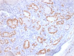
Biotium TTF-1 / NKX2.1 (Thyroid & Lung Epithelial Marker) (rNX2.1/690), CF568 conjugate, 0.1mg/mL
This antibody recognizes a protein of 40 kDa, identified as Thyroid transcription factor-1 (TTF-1). TTF-1 is a member of the NKx2 family of homeodomain transcription factors. It is expressed in epithelial cells of the thyroid gland and the lung. Nuclei from liver, stomach, pancreas, small intestine, colon, kidney, breast, skin, testes, pituitary, prostate, and adrenal glands are unreactive. Anti-TTF-1 is useful in differentiating primary adenocarcinoma of the lung from metastatic carcinomas originating in the breast, mediastinal germ cell tumors, and malignant mesothelioma. It can also be used to differentiate small cell lung carcinoma from lymphoid infiltrates. Loss of TTF-1 expression in non-small cell lung carcinoma has been associated with aggressive behavior of such neoplasms. TTF-1 reactivity is also seen in thyroid malignancies.Primary antibodies are available purified, or with a selection of fluorescent CF Dyes and other labels. CF Dyes offer exceptional brightness
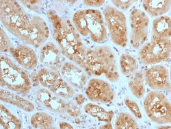
Biotium MTAP (Tumor Suppressor Marker) (MTAP/1813), CF740 conjugate, 0.1mg/mL
This antibody recognizes a protein of 31 kDa, which is identified as MTAP (5'-deoxy-5'-methylthioadenosine phosphorylase). It catalyzes the reversible phosphorolysis of methylthioadenosine, which is important in polyamine metabolism and for the salvage of adenine and methionine. The gene encoding MTAP is linked to the tumor suppressor gene, p16INK4A. Deficient levels of MTAP can occur in cancers primarily through co-deletion of the MTAP gene and the p16INK4A gene. Cells expressing MTAP and possessing adenine salvage pathway activity may be less susceptible to malignancy due to growth-inhibitory actions of agents (e.g. antifolates), whose mechanism of action, in part, involves this de novo purine pathway.Primary antibodies are available purified, or with a selection of fluorescent CF Dyes and other labels. CF Dyes offer exceptional brightness and photostability. Note: Conjugates of blue fluorescent dyes like CF405S and CF405M are not recommended for detecting low abundanc
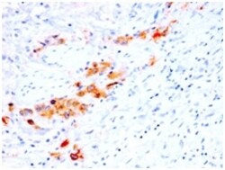
Biotium CD44v4 (Marker of Tumor Metastasis) (rCD44v4/1219), CF568 conjugate, 0.1mg/mL
This antibody recognizes an epitope encoded by exon v4 on the variant portion of human CD44. The CD44 molecule belongs to a family of cellular adhesion molecules found on a wide range of normal and malignant cells in epithelial, mesothelial and hemopoiesis tissues. CD44 is a single gene with 20 exons, of which 10 are normally expressed to encode the basic CD44 (H-CAM) molecule. The additional 10 exons (v1 to v10) are only expressed by alternative splicing of the nuclear RNA. The expression of specific cell adhesion molecule CD44 splice variants has been reported to be associated with metastasis in certain human malignancies.Primary antibodies are available purified, or with a selection of fluorescent CF Dyes and other labels. CF Dyes offer exceptional brightness and photostability. Note: Conjugates of blue fluorescent dyes like CF405S and CF405M are not recommended for detecting low abundance targets, because blue dyes have lower fluorescence and can give higher non-spec
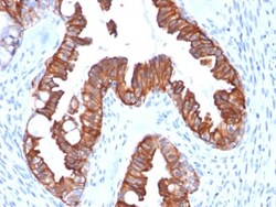
Biotium Cytokeratin 7 (Glandular and Transitional Epithelial Marker) (rOV-TL12/30), 1mg/mL
This antibody recognizes an intermediate filament protein (IFP) of 55 kDa, which is identified as cytokeratin 7. This MAb is highly specific to cytokeratin 7 and shows no cross-reaction with other IFPs. Cytokeratin 7 is a basic cytokeratin, which is found in most glandular and transitional epithelia but not in the stratified squamous epithelia. Keratin 7 is expressed in the epithelial cells of ovary, lung, and breast but not of colon, prostate, or gastrointestinal tract. This MAb is highly useful in distinguishing ovarian carcinomas (keratin 7 ) from colon carcinomas (keratin 7-).Primary antibodies are available purified, or with a selection of fluorescent CF Dyes and other labels. CF Dyes offer exceptional brightness and photostability. Note: Conjugates of blue fluorescent dyes like CF405S and CF405M are not recommended for detecting low abundance targets, because blue dyes have lower fluorescence and can give higher non-specific background than other dye colors.
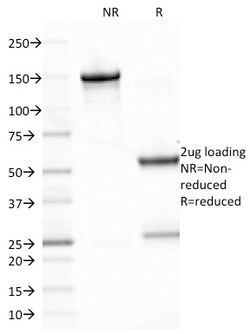
Biotium Epstein-Barr Virus (LMP-1)(CS2), 1mg/mL
This antibody is a mixture of four different monoclonal antibodies. This antibody is specific to 60 kDa latent membrane protein (LMP-1) encoded by the BNLF1 gene of the EBV. Each clone reacts with different epitopes on the hydrophilic C-terminus of the cytoplasmic domain of LMP-1. This antibody stains strongly with EBV-positive lymphoblastoid cell lines and EBV infected B cell immunoblasts in infectious mononucleosis. EBV, also designated human herpesvirus 4 (HHV-4), is a member of the herpesvirus family and is one of the most common human viruses. EBV infects B cells and, though often asymptomatic, it can cause infectious mononucleosis, a disease characterized by fatigue, fever, sore throat and muscle soreness. Primary antibodies are available purified, or with a selection of fluorescent CF Dyes and other labels. CF Dyes offer exceptional brightness and photostability. Note: Conjugates of blue fluorescent dyes like CF405S and CF405M are not recommended for detecting low
Biotium MUC1 / CA15-3 / EMA / CD227 (Epithelial Marker)(MUC1/1887R), 1mg/mL
This MAb reacts with MUC1, a large transmembrane glycoprotein expressed on the ductal surface of normal glandular epithelia. It is used as tracer agent in CA15.3 assays. The extracellular domain of MUC1 largely consists of a highly conserved, O-glycosylated 20 amino acids tandem repeat which can occur 30-100 times per molecule depending on the length of the allele involved. In the vast majority of human carcinomas this protein is up-regulated and poorly glycosylated and appears on the cell surface in a non-polarized fashion. The dominant epitope of this MAb involves both amino acids as well as sugar moieties. Its epitope is destroyed by desialylation i.e. treatment with Neuraminidase.Primary antibodies are available purified, or with a selection of fluorescent CF Dyes and other labels. CF Dyes offer exceptional brightness and photostability. Note: Conjugates of blue fluorescent dyes like CF405S and CF405M are not recommended for detecting low abundance targets, because b
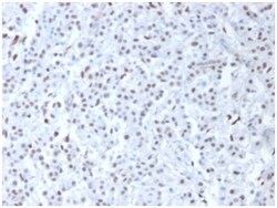
Biotium Wilm's Tumor 1 (WT1) (Wilm's Tumor & Mesothelial Marker), 0.2mg/mL
Recognizes a 47-55 kDa-tumor suppressor protein, identified as Wilm's Tumor (WT1) protein. The antibody reacts with all isoforms of the full-length WT1 and also identifies WT1 lacking exon 2-encoded amino acids, frequently found in subsets of sporadic Wilms tumor and mesothelioma. WT1 protein has been identified in proliferative mesothelial cells, malignant mesothelioma, ovarian carcinoma, gonadoblastoma, nephroblastoma, and desmoplastic small round cell tumor. Lung adenocarcinomas rarely stain positive with this antibody. WT1 protein expression in mesothelial cells has become a reliable marker for the diagnosis of mesotheliomas. Primary antibodies are available purified, or with a selection of fluorescent CF Dyes and other labels. CF Dyes offer exceptional brightness and photostability. Note: Conjugates of blue fluorescent dyes like CF405S and CF405M are not recommended for detecting low abundance targets, because blue dyes have lower fluorescence and can give higher no
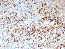
Biotium HSV1 (Herpes Simplex Virus Type I)(N/A), 0.2mg/mL
The antibody reacts with HSV type 1 specific antigen. It is suitable for detection of HSV in human cellular material obtained from superficial lesions or biopsies and for the early identification of HSV in infected tissue cultures. The herpes simplex virus (HSV) (also known as cold sore, night fever or fever blister) is a virus that causes a contagious disease. There are two main types of Herpes Simplex Virus (HSV), 1 and 2. The HSV-1 strain generally appears in the orafacial organs. HSV2 usually resides in the sacral ganglion at the base of the spine. All herpes viruses are morphologically identical: they have a large double-stranded DNA genome and the virion consists of an icosahedral nucleo-capsid, which is surrounded by a lipid bilayer envelope. Primary antibodies are available purified, or with a selection of fluorescent CF Dyes and other labels. CF Dyes offer exceptional brightness and photostability. Note: Conjugates of blue fluorescent dyes like CF405S and CF405M
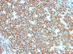
Biotium CD20 / MS4A1 (B-Cell Marker) (rIGEL/773), 0.2mg/mL
This antibody recognizes a protein of 30-33 kDa, which is identified as CD20. It is a non-Ig differentiation antigen of B-cells and its expression is restricted to normal and neoplastic B-cells, being absent from all other leukocytes and tissues. CD20 is expressed by pre B-cells and persists during all stages of B-cell maturation but is lost upon terminal differentiation into plasma cells. This MAb can be used for immunophenotyping of leukemia and malignant cells, B lymphocyte detection in peripheral blood and B cell localization in tissues. It reacts with the majority of B-cells present in peripheral blood and lymphoid tissues and their derived lymphomas. In lymphoid tissue, germinal center blasts and B-immunoblasts are particularly reactive. It is a reliable antibody for ascribing a B-cell phenotype in known lymphoid tissues. Rarely, CD20-positive T-cell lymphomas have been reported. Reactivity has also been noted with Reed-Sternberg cells in cases of Hodgkin's disease, par
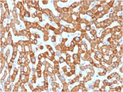
Biotium Prohibitin (Mitochondrial Marker)(PHB/1881), 0.2mg/mL
Recognizes a protein of 30 kDa which is identified as Prohibitin, an evolutionarily conserved protein with homologues found in yeast to man. It is located in the inner membrane of mitochondria. Although prohibitin mRNA and protein expression occurs throughout the cell cycle, maximum levels are detected during the G1/S phase transition and minimum levels are seen in S phase and the G2/mitosis boundary. Prohibitin is located exclusively in the mitochondria with the highest concentration on the inner membrane. Prohibitin is an ideal mitochondrial marker. It shows antiproliferative activity and has been proposed to play a role in normal cell cycle regulation, replicative senescence, cellular immortalization, and tumor suppression. Primary antibodies are available purified, or with a selection of fluorescent CF Dyes and other labels. CF Dyes offer exceptional brightness and photostability. Note: Conjugates of blue fluorescent dyes like CF405S and CF405M are not recommended fo
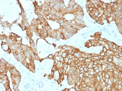
Biotium Cytokeratin 19 (Pancreatic Stem Cell Marker) (rKRT19/800), Biotin conjugate, 0.1mg/mL
This antibody recognizes a protein of 40 kDa, identified as cytokeratin-19 (CK19), which is expressed in sweat gland, mammary gland ductal and secretory cells, bile ducts, gastrointestinal tract, bladder urothelium, oral epithelia, esophagus, and ectocervical epithelium. Anti-CK19 reacts with a wide variety of epithelial malignancies including adenocarcinomas of the colon, stomach, pancreas, biliary tract, liver, and breast. Perhaps the most useful application is the identification of thyroid carcinoma of the papillary type, although 50%-60% of follicular carcinomas are also labeled. Anti-CK19 is a useful marker for detection of tumor cells in lymph nodes, peripheral blood, bone marrow and breast cancer.Primary antibodies are available purified, or with a selection of fluorescent CF Dyes and other labels. CF Dyes offer exceptional brightness and photostability. Note: Conjugates of blue fluorescent dyes like CF405S and CF405M are not recommended for detecting low abundanc